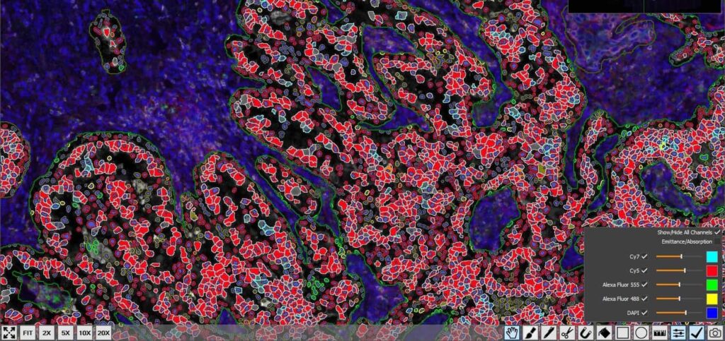
The Use of Image Fusing in the Deployment of a 7-plex Immunofluorescent Assay
27 April 2020 | In lieu of our participation in the now cancelled 2020 AACR Conference, Indica Labs are pleased to bring you webinar presentations from two of our esteemed customers in oncology research and drug discovery, David Krull from GlaxoSmithKline and Dr. David Reiss from Bristol Meyers Squibb, who will present on April 27th and 28th, respectively. Please join us to hear about how Indica Labs image analysis software and pharma services are being used within their organizations to advance oncology drug development.
The Use of Image Fusing in the Deployment of a 7-plex Immunofluorescent Assay Read More »